
Figure 6:
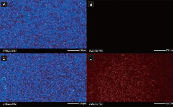
Fluorescence microscopy images of (A) C. albicans cells and (B) C. albicans dead cells in biofilm treated with green laser alone; (C) C. albicans cells and (D) C. albicans dead cells in biofilm treated with Erythrosine, green laser and KP; scale bar of 250 rm, magnification 10×. Panel A and C, staining with calcofluor white; panel B and D, staining with propidium iodide. The absence of candidacidal effect for green laser alone is visible in panel B as absence of propidium iodide (red) cells.