
Figure 2.
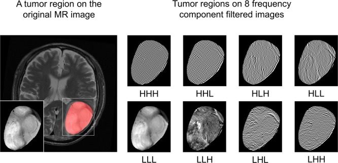
Transverse images of a tumor on original magnetic resonance (MR) image (T2-weighted MR image (T2WI)) and on eight frequency component-filtered images to which a three-dimensional (3D) Coiflet wavelet transform had been applied.
Official websites use .gov
A
.gov website belongs to an official
government organization in the United States.
Secure .gov websites use HTTPS
A lock (
) or https:// means you've safely
connected to the .gov website. Share sensitive
information only on official, secure websites.
Transverse images of a tumor on original magnetic resonance (MR) image (T2-weighted MR image (T2WI)) and on eight frequency component-filtered images to which a three-dimensional (3D) Coiflet wavelet transform had been applied.