Figure 4.
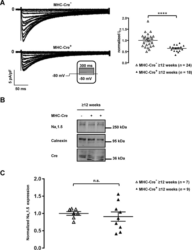
Decreased ICaL and unaltered Nav1.5 expression in MHC-Cre+ mouse cardiomyocytes and mouse hearts respectively. (A) Representative traces of ICaL recorded in isolated cardiomyocytes (left panel) and quantification of ICaL (right panel) shown a significant downregulation of the calcium current in MHC-Cre+ cardiomyocytes compared to the control (MHC-Cre−) from ≥12-week-old mice. ****p < 0.0001. (B) Cropped western blots showing Nav1.5 expression in heart lysates from MHC-Cre+ and MHC-Cre− ≥12-week-old mice. The bottom panel shows the quantification of Nav1.5 expression normalized to calnexin expression from 7 to 9 western blots. n.s. indicates a non-statistically significant difference.

