Figure 7.
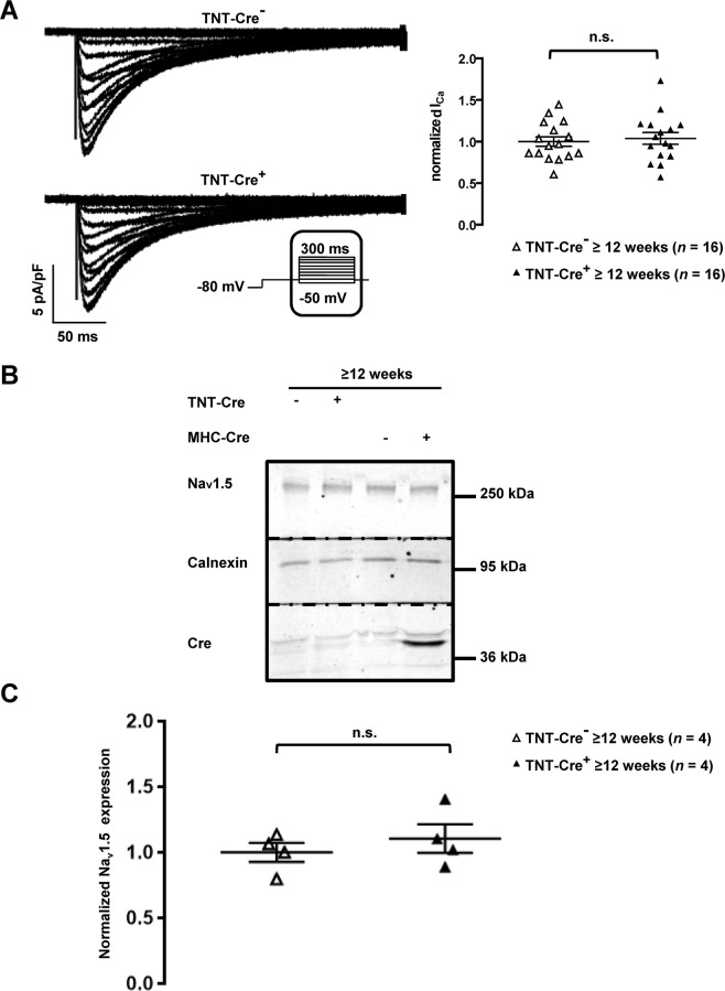
ICaL and Nav1.5 expression are unaltered in TNT-Cre+ mouse cardiomyocytes and mouse hearts. (A) Representative traces of ICaL recorded in isolated cardiomyocytes (left panel) and quantification of ICaL (right panel) show that the calcium current is not changed in TNT-Cre+ cardiomyocytes compared to the control (TNT-Cre−) from ≥12-week-old mice. n.s. indicates a non-statistically significant difference. (B) Cropped western blots showing Nav1.5 expression in heart lysates from ≥12-week-old MHC-Cre+, MHC-Cre−, TNT-Cre−, and TNT-Cre+ mice. (C) Nav1.5 expression quantification of 4 western blots normalized to calnexin band intensity. n.s. indicates a non-statistically significant difference.

