Figure 1.
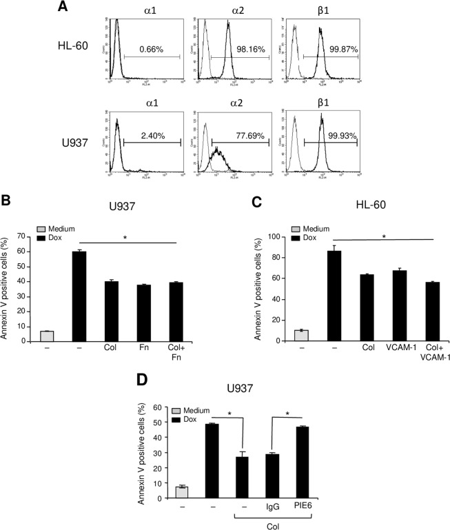
Collagen via α2β1 integrin protects AML cell lines from doxorubicin-induced apoptosis. (A) Flow cytometry analysis of α1, α2 and β1 integrin subunits expression on HL-60 and U937 cells. (B,C) Collagen reduces doxorubicin-induced apoptosis of U937 and HL-60 cells. The cells were cultured on BSA (–), collagen (Col), fibronectin (Fn) or on VCAM-1 as indicated for 2 h. Cells in suspension were washed and adherent cells were treated with 1 μM doxorubicin (Dox) for 24 h. Apoptosis was determined by annexin V staining and flow cytometry analysis. The results represent mean values ± SD from three independent experiments. *P < 0.05 between doxorubicin-treated samples cultured on collagen, fibronectin or VCAM-1 and doxorubicin-treated samples cultured on BSA (–). (D) α2 integrin blockade reverses the collagen protective effect. U937 cells were pretreated with 10 μg/ml of anti-α2 blocking antibody (PIE6) or with isotypic control IgG for 1 h before their culture on collagen. The cells were then treated with doxorubicin and apoptosis was determined by annexin V staining and flow cytometry analysis. The results represent mean values ± SD from three independent experiments. *P < 0.05.

