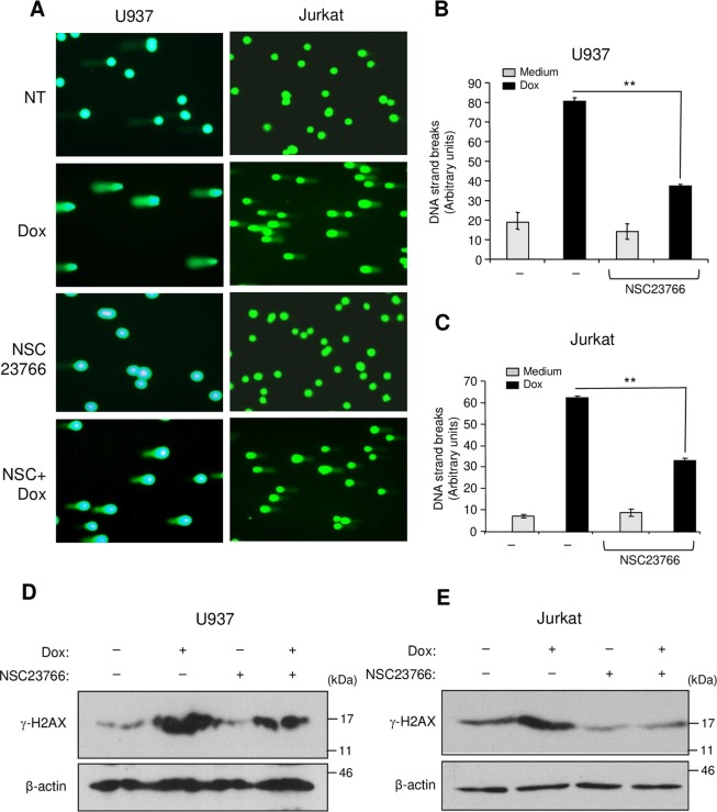
Figure 6.
Rac1 inhibition reduces DNA damage intensity and H2AX phosphorylation induced by doxorubicin. (A–C) The cells were treated or not with doxorubicin (Dox) for 6 h in the presence or absence of the Rac1 inhibitor NSC23766 (NSC). Alkaline comet assay was performed and stained nucleoids were visualized by epifluorescence microscopy using FITC filter. (A) Representative fields corresponding to each treatment were photographed. (B,C) The intensity of DNA strand breaks in U937 and Jurkat cells was quantified using visual scoring as described under “Experimental procedures section”. The results represent mean values ± SD obtained from three independent experiments. **P < 0.01. (D,E) The cells were treated with doxorubicin in the presence or absence of NSC23766 as described above and the levels of phosphorylated H2AX (γ-H2AX) were determined by immunoblot analysis. The β-Actin blot was used as a loading control. Blots are representative of three independent experiments.

