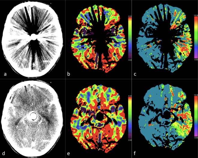
Figure 4.
CT perfusion study of a 60-year-old female patient after coiling of a ruptured basilar tip aneurysm. The upper panel (a–c) show images before, and the lower panel (d–f) show images after iMAR application. Obvious metal artifact reduction was apparent after iMAR application, which improved the detection of MTT prolongation in the left temporal lobe (c,f) and increased rater confidence. Rater confidence in analysing the CBV (b,e), which showed no asymmetry even after artifact reduction, was also increased. Note: A hypodense ring artifact surrounded by a hyperdense ring was observed around the metal in the source images processed with iMAR (d).

