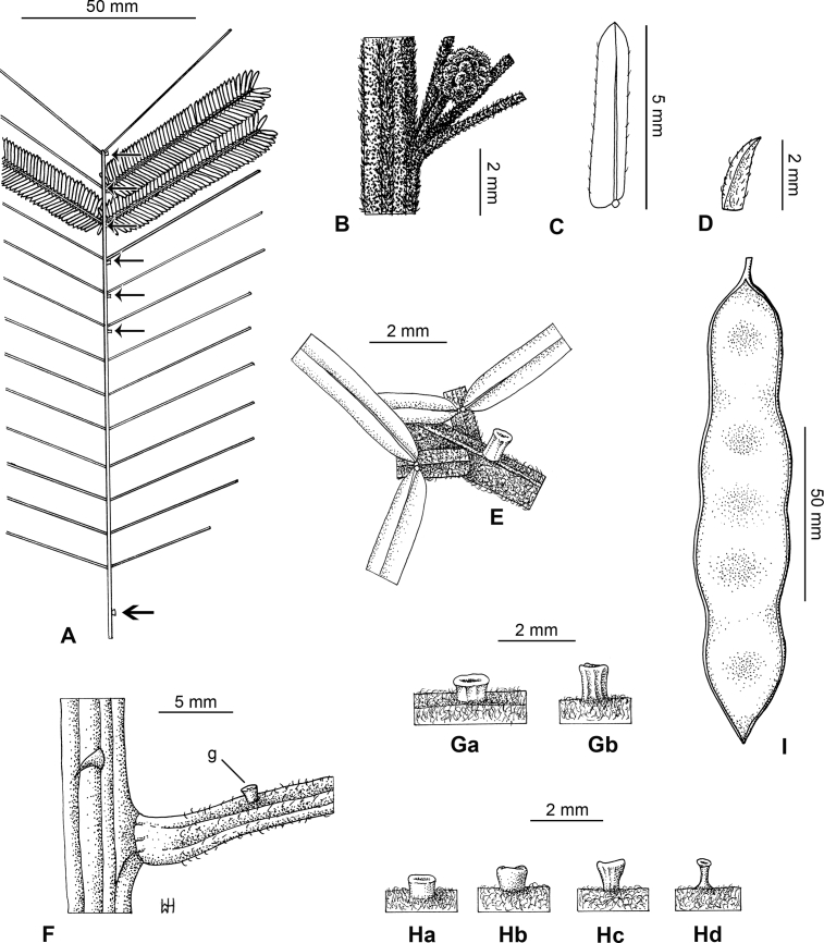
Fig. 47.
Senegalia stipitata. A – Leaf showing position of petiole gland (thick arrow) and numerous rachis glands (thin arrows). B – Inflorescence bud at node, showing dark-colouring due to dense indumentum of resin hairs. C – Leaflet (lower surface) showing main situated slightly towards and parallel with upper margin near base of leaflet. D – Stipule small. E − Rachis gland (stipitate) at base of pinnae. F – Node showing small, stipitate petiole gland position. G – Rachis glands (stipitate) showing variation. H – Petiole glands (stipitate) showing variation (Hd = calicioid; Hc & Hd – note variation from the one specimen). I – Pod. Vouchers: Sino-Vietnam Expedition 1603 (A, C, E, F, Gb & Ha); P.I. Mao 3922 (B & D); A.J.B. Chevalier 37410 (Ga); Ban, Phuong, Khoi, Binh & Bach 2067 (I); Anonymous (Hb –IBSC 680597); K.H. Cai 1265 (Hc & Hd). Drawn by Waiwai Hove.

