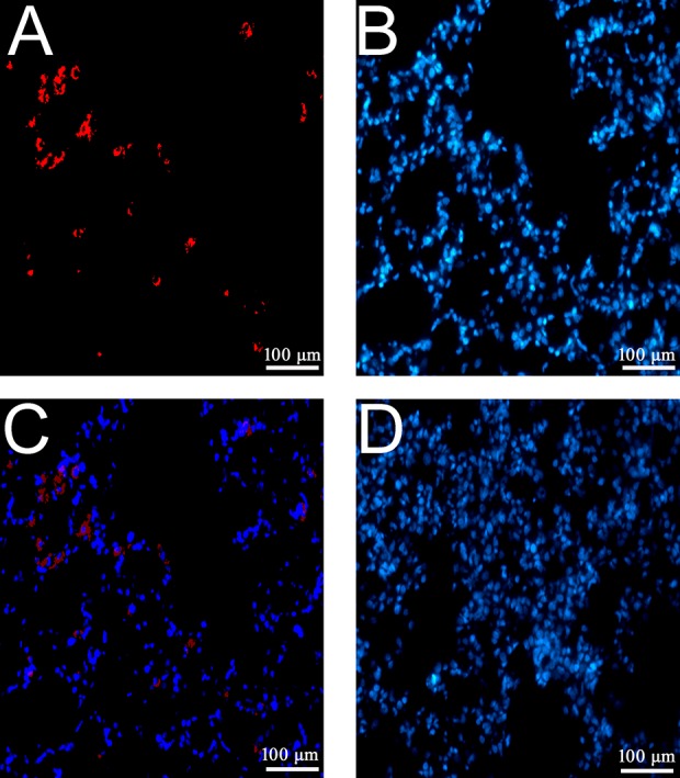
Figure 2.

Tracking EPCs following the transplantation in lung tissue. EPCs transplanted to lung tissues at 24 h after injection were detected and identified by acetyl-LDL immunofluorescence staining. The positive acetyl-LDL stain presented as red (A), and the nuclei was visualized by DAPI (blue, B). The merged image is presented in C (200 × magnification). The DAPI stain in the tissue of rats in the V group is presented in D. Abbreviations: DAPI, 4-,6-diamidino-2-phenylindole; EPCs, endothelial progenitor cells; LDL, low density lipoprotein. Each testing group contained eight rats.
