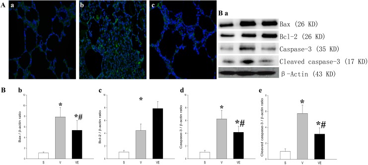
Figure 6.
EPC transplantation mitigated the ventilation-induced apoptotic damage in lung
tissue. The representative results of apoptosis in lung tissue in the S group (Aa), V
group (Ab), and EPC transplantation (VE group; Ac) were detected by TUNEL staining
using fluorescence microscopy (A, 400 × magnification). TUNEL-positive cells were
stained green and the nucleus was stained blue with DAPI. The representative results
of the protein expression of Bax (first panel of Ba and Bb), Bcl-2 (second panel of Ba
and Bc), native caspase-3 (third panel of Ba and Bd), and cleaved caspase-3 (fourth
panel of Ba and Be) in lung tissue in the S group (left bands of Ba,  ), V group (middle bands of Ba;
 ) and VE group
(right bands of Ba;
  )
were detected by Western blot. The expression of the corresponding proteins was
evaluated by densitometry analysis, and presented in Bb (Bax), Bc (Bcl-2), Bd (native
caspase 3), and Be (cleaved caspase 3). Data in each group were calculated from three
independent tests. *p < 0.05 vs. the S group;
#
p < 0.05 vs. the V group. Abbreviations: EPCs,
endothelial progenitor cells; S group, sham; TUNEL, terminal deoxynucleotidyl
transferase dUTP nick end labeling; V group, VILI; VE group, VILI with EPC
transplantation; VILI, ventilator-induced lung injury.

