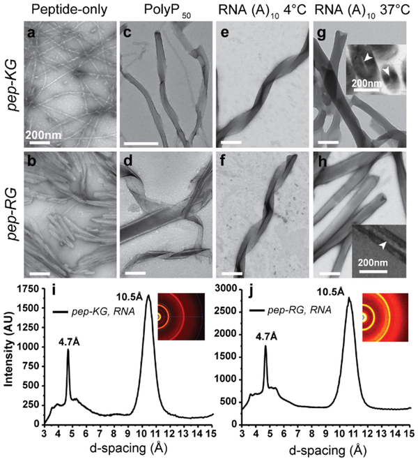
Figure 3. RNA/Peptide co-assembly.
(a/b) TEM micrographs of pep-KG and pep-RG self-assemblies. (c/d) PolyP50 templated assembly of pep-KG and pep-RG. (e/f) RNA (A10)-templated assembly of pep-KG and pep-RG at 4°C. (g/h) At 37°C, the ribbons fuse to form thick-walled nanotubes; TEM Insets show nanotube cross-sections. (i/j) Powder x-ray diffraction of peptide/RNA co-assemblies. Pep-KG refers to Ac-KLVIIAG-NH2 and pep-RG to Ac-RLVIIAG-NH2. Arrowheads indicate hollow nanotube interior. Scale bars are 200nm.

