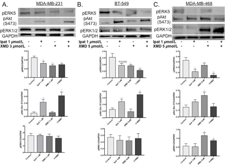
Figure 2. Analysis of ERK5, Akt and ERK1/2 activation in TNBC cells.
MDA-MB-231 (A), BT-549 (B), and MDA-MB-468 (C) cells were treated with Akt inhibitor, Ipatasertib, and ERK5 inhibitor, XMD8-92, for 24 hours under 5% FBS stimulation. Western blot analysis was performed on cellular lysates. GAPDH was used as a loading control. *P<0.05 and **P<0.01 vs control.

