Fig. 7.
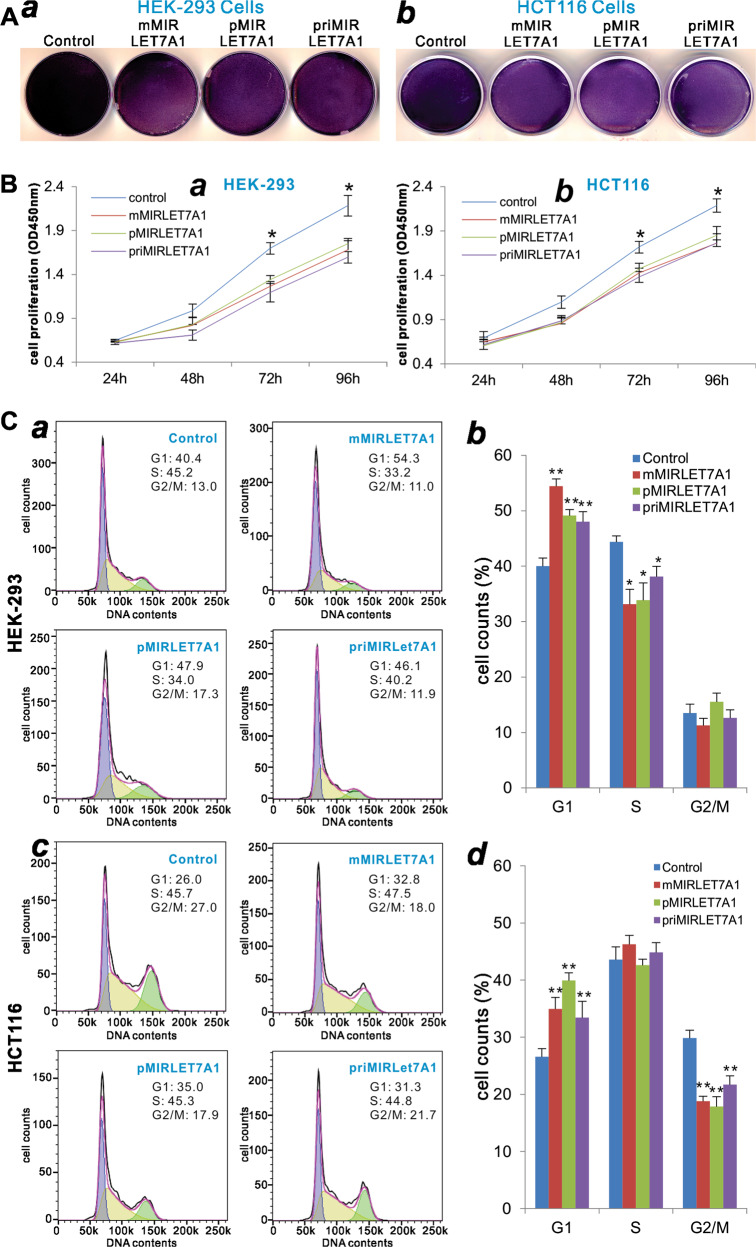
Comparison of the let-7a-1-inhibited cell proliferative activity mediated by the three let-7a-1 expression systems. a The crystal violet assay. Subconfluent HEK-293 (a) and HCT116 (b) cells were transfected with the indicated let-7a-1 expression plasmids or vector control. Cells were fixed for Crystal violet staining at 3 days after transfection. Assays were done in triplicate, and representative results are shown. b The WST-1 cell proliferation assay. Subconfluent HEK-293 (a) and HCT116 (b) cells were transfected with the indicated let-7a-1 expression plasmids or vector control. At the indicated time points, WST-1 substrate was added to each well and incubated for 2 h, followed by measuring the absorbance at 450 nm. Assays were done in triplicate. *p < 0.05, **p < 0.01 compared with the vector control group. c Cell cycle analysis. Subconfluent HEK-293 (a, b) and HCT116 (c, d) cells were transfected with the indicated let-7a-1 expression plasmids or vector control. At 48 h after transfection, cells were harvested and subjected to cell cycle analysis. Assays were done in triplicate, and representative results are shown (a, c). Quantitative analyses were carried out to determine the % cell counts in various phases (b, d). *p < 0.05 and **p < 0.01 compared with the vector control group

