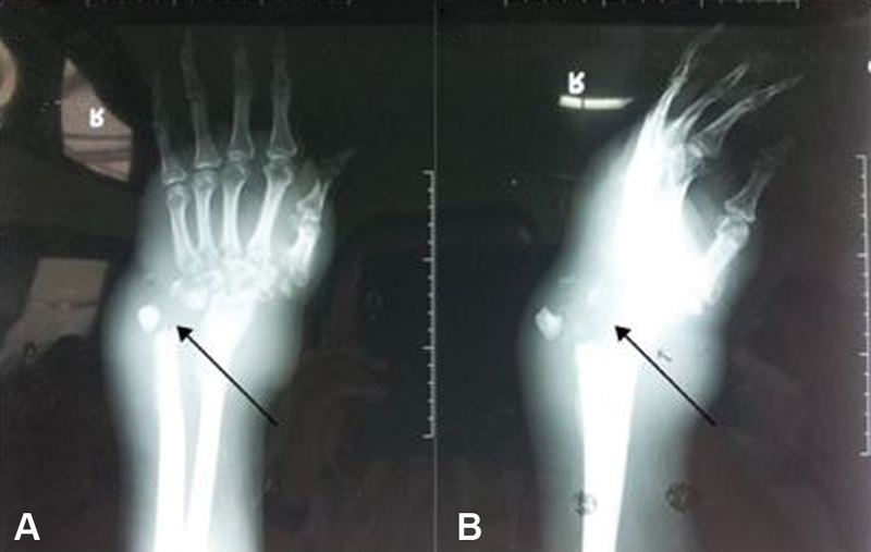
Fig. 5.

A 44-year-old male with tuberculous osteomyelitis on his right wrist. Anteroposterior ( A ) and lateral ( B ) views of plain radiograph of the right wrist revealed a lytic lesion on the distal radius, ulna, and carpal bone, with soft tissue swelling.
