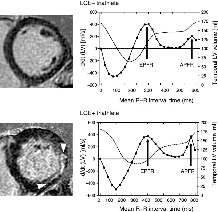
Figure 1.
Left ventricular (LV) volume (dashed line) and differentiated LV time-volume (solid line) curves analysed using cine cardiac magnetic resonance in a LGE– and LGE + triathlete. The differentiated time-volume curve is characterised by two diastolic peaks including the early peak filling rate (EPFR) and the atrial peak filling rate (APFR). The LGE– triathlete had normal peak filling rates with high EPFR and low APFR, whereas the LGE + triathlete had an increased APFR. LGE: late gadolinium enhancement.

