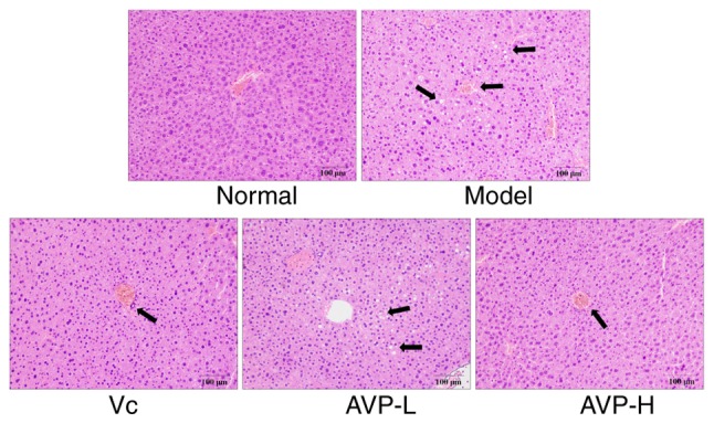
Figure 3.

Hematoxylin and eosin staining of mouse liver. Magnification, ×100. Vc, mice treated with 100 mg/kg Vc; AVP-L, mice treated with a low concentration (50 mg/kg) of AVP; AVP-H, mice treated with a high concentration (100 mg/kg) of AVP. AVP, Apocynum venetum polyphenol extract; Vc, vitamin C. Arrow indicates inflammed tissue.
