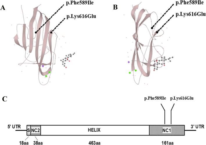
Fig. 4.
Protein modeling of type X collagen (α1) NC1 domain and stylized structure of collagen X. a and b As illustrated in the ribbon protein model, both of the novel mutations are located in the NC1 domain of type X collagen (α1). One of the substitutions (p.Phe589Ile) affects a hydrophobic area and the other (p.Lys616Glu) is predicted to affect the surface of the assembled trimer. c The stylized structure of type X collagen (α1) is composed of a 18 amino acid signal peptide (S) and a 463 amino acid collagenous domain (HELIX) flanked by a 38-residue NC2 domain and a 161-residue NC1 domain. Furthermore, changes in the present study and most previous variants (Additional file 1) are located in NC1 domain

