Fig. 1.
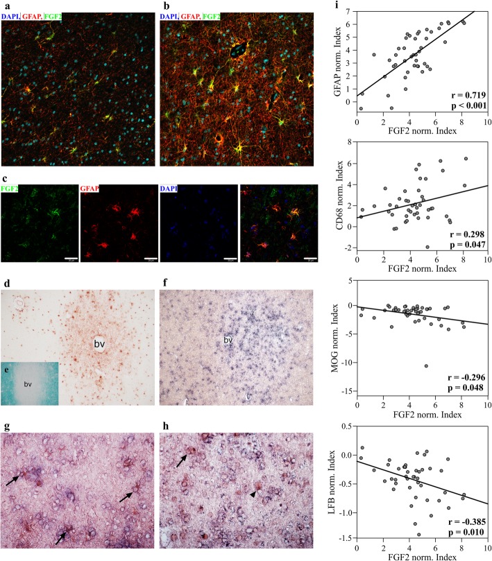
FGF2 is up-regulated in astrocytes of subcortical multiple sclerosis lesions. Fluorescence immunostaining for FGF2 and GFAP of NAWM (a) and active multiple sclerosis lesion (b) show strong FGF2 expression in the lesion. FGF2 expression co-localizes with GFAP (c). This finding is supported by immunohistochemistry of FGF2 expression in multiple sclerosis lesion (d). Demyelinating lesion is indicated by reduced Fast Luxol Blue staining around the blood vessel (e). In situ hybridization depicts Fgf2 transcripts within active lesion (f) mainly in GFAP-positive cells (g, arrows), and only rarely and weakly in Olig2-positive cells (h, arrow). Most of the Olig2-positive cells do not express FGF2 (h, arrowhead). Note, ISH is weaker after immunohistochemistry. Magnifications a, b, g and h: 40x; d-f: 10x; bv: blood vessel, c: scale bars represent 20 μm. i Quantification of FGF2 intensity in correlation to GFAP, MOG, CD68 and LFB shows positive correlation with GFAP and inflammation (CD68) and negative correlation with myelin reactivity (LFB and MOG). Ten randomly taken images from at least 3 areas of interest per multiple sclerosis patient (n = 7 patients) were analyzed and normalized to the corresponding values in the NAWM of the patient. Data was log2 transformed and Pearson correlation over all cases was calculated

