Figure 4.
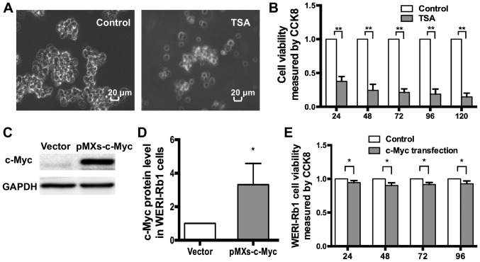
Exogenous c-myc affects the viability of WERI-Rb1 cells. (A) Morphological changes in WERI-Rb1 cells treated with TSA (250 nM) for 72 h. (B) CCK-8 assays indicated that TSA significantly decreased the viability of WERI-Rb1 cells compared with the negative controls. **P<0.01. (C) Western blotting showing the expression of c-Myc in WERI-Rb1 cells after transfection. (D) Relative expression of c-Myc in WERI-Rb1 cells was quantified using densitometry; the data are presented as histograms. *P<0.05 vs. vector. (E) CCK-8 assays indicated that the viability of WERI-Rb1 cells was reduced following c-myc transfection, compared with the controls. All results were confirmed in triplicate. *P<0.05. TSA, trichostatin A; CCK-8, Cell Counting Kit-8.

