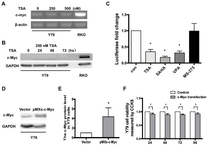
Figure 5.
c-Myc is not upregulated by HDAC inhibitors in RB Y79 cells. (A) RT-PCR analysis indicated that c-myc expression was not induced by treatment with 250 or 500 nM TSA, in Y79 cells. (B) Western blot analysis of c-Myc in Y79 cells following TSA treatment. GAPDH was used as a reference gene. (C) Y79 cells were transfected with the c-myc promoter reporter and then treated with 250 nM TSA, 10 µM SAHA, 10 mM VPA or 5 µM MS-275 for 24 h. Levels of luciferase activity were normalized to those of Renilla luciferase (control, 1; TSA, 0.35±0.07; SAHA, 0.18±0.04; VPA, 0.31±0.59; MS-275, 0.99±0.24; n=3 for each group) *P<0.05 vs. control. (D) Expression level of c-Myc in Y79 cells after c-Myc transfection. (E) Relative expression of c-Myc in Y79 cells was quantified using densitometry, and the data are presented as histograms. *P<0.05 vs. vector. (F) CCK-8 assays indicated that the viability of Y79 cells may also be reduced by exogenous c-Myc, similar to WERI-Rb1 cells. All the results were confirmed in triplicate. *P<0.05. RT-PCR, reverse transcription PCR; TSA, trichostatin A; SAHA, vorinostat; VPA, valproic acid sodium salt; MS-275, entinostat; con, control.

