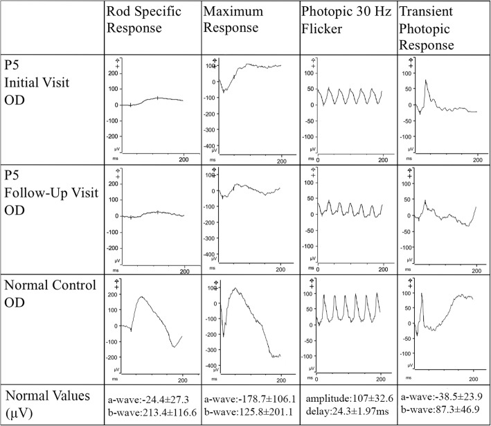
Fig. 4.
Rod Cone Dysfunction in Full Field Electroretinogram Findings of Patient 5. Full-field electroretinogram findings of the right eye of P5 across two visits separated by 2 years demonstrated a slow decrease in both scotopic rod-specific and photopic 30 Hz flicker suggestive of slow disease progression. Normal values were demonstrated through an age-matched control patient

