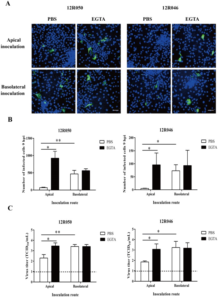
Figure 3.
Rotavirus preferentially infects the basolateral surface of enterocytes and disruption of ICJ overcomes the restriction of rotavirus infection at the apical surface. A Representative confocal images of rotavirus infection (green) in enterocytes. Scale bar: 50 µm. To compare epithelial cell susceptibility to rotavirus, cells were exposed at either the apical surface or the basolateral surface to rotavirus 12R050 (G5P[7]) and 12R046 (G9P[23]). B The total number of infected cells per well was counted for each condition. C The virus titer was determined in supernatant for each condition. Data are expressed as the mean ± SD of the results of three separate experiments. Statistically significant differences are indicated with one asterisk (p < 0.05) and two asterisks (p < 0.01).

