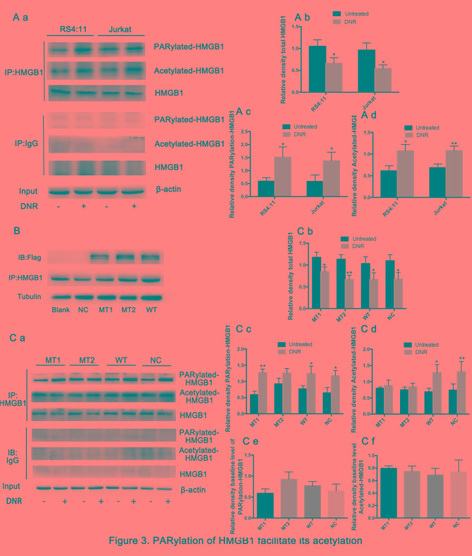
Figure 3.
Poly (ADP-ribosylation) of HMGB1 facilitates its acetylation. (A) Cell lysates were immunoprecipitated with an HMGB1 antibody, followed by western blot analysis. The acetylation or poly (ADP-ribosylation) levels were measured using antibodies specific for acetylated lysine or poly (ADP-ribosylation). β-actin was used as a loading control. Quantified data are presented (HMGB1/β-actin, PARylation-HMGB1 or Acetylated-HMGB1/HMGB1/β-actin). (a Western blot diagram of RS4:11 and Jurkat cells. (b) Total HMGB1 quantitative data of RS4:11 and Jurkat cells. (c) PARylation-HMGB1 quantitative data of RS4:11 and Jurkat cells. (d) Aceytlation-HMGB1 quantitative data of RS4:11 and Jurkat cells. (B) HMGB1MT1, HMGB1MT2 and HMGB1WT cells were successfully constructed. The lysine residues at amino acids 28, 29, 30, 180, 182, 183, 184 and 185 of HMGB1 in HMGB1MT1 cells were mutated to alanine, and the glutamate residues at 40, 47 and 179 of HMGB1 in HMGB1MT2 cells were mutated to alanine. The whole protein of HMGB1MT1, HMGB1MT2, HMGB1WT, HMGB1NC and Jurkat cells were subjected to immunoprecipitation with anti-HMGB1 antibodies and then subjected to western blotting to detect the expression of lentivirus with anti-Flag antibodies. Tubulin was used as a loading control. (C) Cell lysates were subjected to a pull-down assay with an HMGB1 antibody and immunoblotted with anti-acetylated lysine, anti-poly (ADP-ribosylation) and anti-HMGB1 antibodies. β-actin was used as a loading control. Quantified data are presented (HMGB1/β-actin, PARylation-HMGB1 or Acetylated-HMGB1/HMGB1/β-actin). Data are the mean ± standard deviation of three independent experiments. (a) Western blot diagram of MT1, MT2, WT and NC cells. (b) Total HMGB1 quantitative data of MT1, MT2, WT and NC cells. (c) PARylation-HMGB1 quantitative data of MT1, MT2, WT and NC cells. (d) Aceytlation-HMGB1 quantitative data of MT1, MT2, WT and NC cells. *P<0.05, **P<0.01, Fig. 3Cb-d compared with the untreated group and Fig. 3Ce and f compared with the NC group. HMGB1, high mobility group box protein 1; DNR, daunorubicin; MT, mutant type; WT, wild type; NC, normal control; UT, untreated group.

