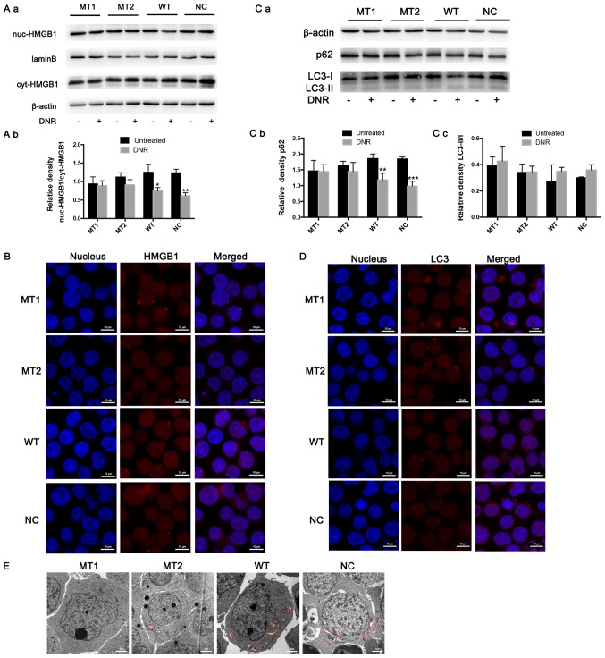
Figure 4.
HMGB1 translocation is associated with chemotherapeutic drug-induced autophagy. (A) Cell lysates were separated into cytosolic and nuclear fractions. Cytosolic and nuclear HMGB1 were assayed via western blotting. β-actin was used as a loading control to detect the expression of cytoplasmic protein, while laminB was used to detect the expression of nuclear protein. Quantified data are presented [(nuc-HMGB1/laminB)/(cyt-HMGB1/β-actin)]. (a) Western blot diagram of MT1, MT2, WT and NC cells. (b) nuc-HMGB1/cyt-HMGB1 quantitative data of J MT1, MT2, WT and NC cells. (B) Intracellular HMGB1 was stained via indirect immunofluorescence and analysed under a confocal microscope to detect the location of HMGB1 (HMGB1, Cy3 staining; nucleus, DAPI staining). (C) The cell lysates were subjected to western blotting to detect LC3-II/I and p62 expression. β-actin was used as a loading control. Quantified data are presented (p62 or LC3-II/I/β-actin). (a) Western blot diagram of MT1, MT2, WT and NC cells. (b) p62 quantitative data of MT1, MT2, WT and NC cells. (c) LC3II/I quantitative data of MT1, MT2, WT and NC cells. (D) LC3 was stained via indirect immunofluorescence and analysed under a confocal microscope to measure the LC3 puncta (LC3, staining with Cy3; nucleus, staining with DAPI). (E) Cells were subjected to transmission electron microscopy to observe autophagosome-like structures (indicated by red circles). Data are the mean ± standard deviation of three independent experiments. *P<0.05, **P<0.01, ***P<0.001 compared with the untreated group. HMGB1, high mobility group box protein 1; MT, mutant type; WT, wild type; NC, normal control; UT, untreated group; DNR, daunorubicin.

