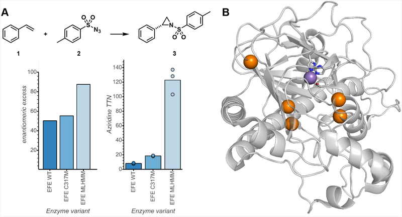
Figure 2.
Directed evolution of PsEFE for aziridination. (A) Evolutionary lineage. Reactions were performed in triplicate anaerobically with acetate and quantified by analytical HPLC-UV. Full experimental details are given in the Supporting Information. (B) Structural representation of PsEFE with mutated sites highlighted in orange; metal-coordinating residues H189, D191, and H268 are represented in sticks and Mn (the metal with which the protein was crystallized) is represented as a purple sphere (PDB ID: 6CBA).

