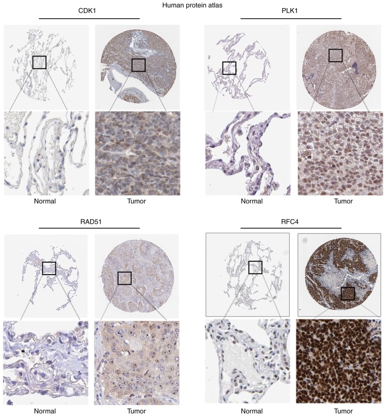
Figure 5.
Hub gene protein expression in human NSCLC specimens was determined from The Human Protein Atlas. Representative IHC images of hub gene protein expression in NSCLC tissues and normal lung tissues. Each lower panel is a enlargement of the outlined area in the top panel in its respective column in the same sample. The IHC analysis demonstrated that CDK1 was highly expressed in NSCLC compared with that in normal lung samples, which was also true for PLK1, RAD51 and RFC4. The IHC images were downloaded from The Human Protein Atlas. NSCLC, non-small cell lung cancer; IHC, immunohistochemistry.

