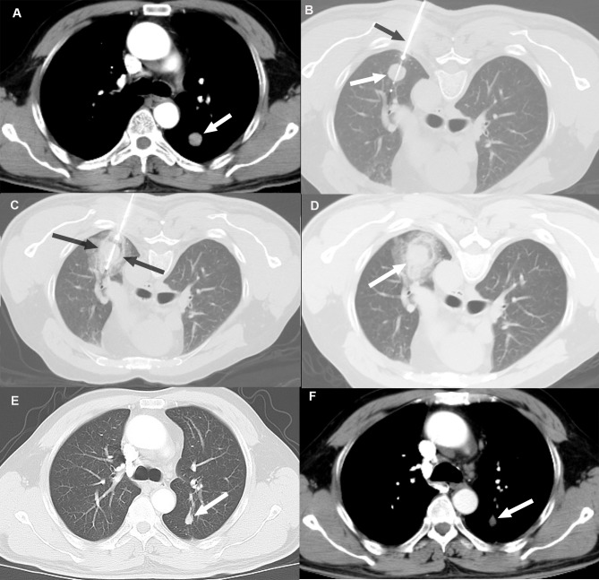
Figure 1.
Local disease control at six months post-CA in a 48-year old male patient with a 14.8-mm left lower lobe primary tumor. (A) Pre-ablation contrast-enhanced axial CT images in soft-tissue windows showing a primary tumor (white arrow). (B and C) Axial CT images during the CA procedure showing (B) cryoprobe (black arrow) positioned within the tumor (white arrow) and (C) excellent coverage of tumor with the ice ball (between black arrows) following freezing. (D) Images captured immediately post-CA. (E and F) Contrast-enhanced follow-up CT images in (E) lung and (F) soft-tissue windows 6 months after CA demonstrating tumor size reduction without evidence of enhancement (white arrow). CA, cryoablation.

