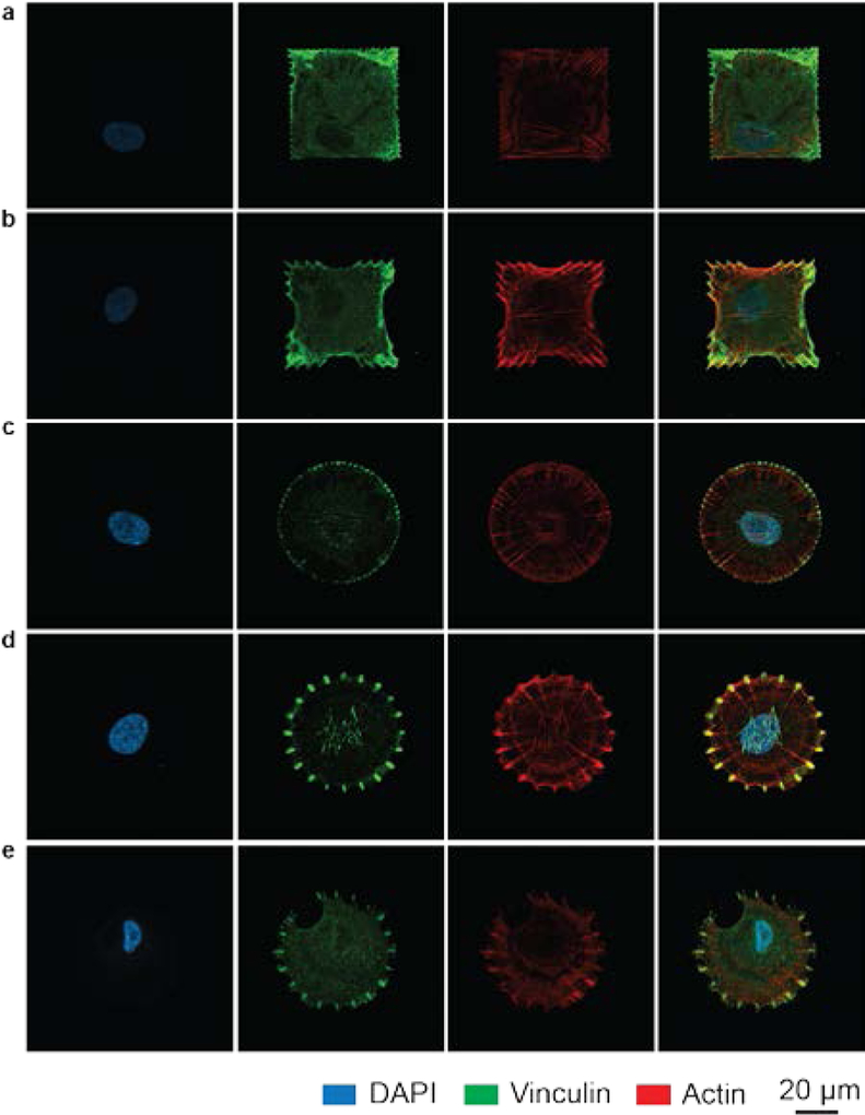
Figure 3. Fluorescence micrographs of the focal adhesions and actin cytoskeleton within cells on patterns.
a-e, Representative confocal images of single cells grown on different patterns: square dot matrix (a), anisotropic square (b), dot matrix circles (c), 20-point circles (d), and hybrid circles (e). The nucleus (Panel 1), focal adhesions (vinculin, panel 2), and actin cytoskeleton (panel 3) are labeled within each cell. Panel 4 shows the overlay of the different structures.

