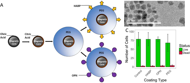
Figure 1:

(A) Schematic of particle fabrication and functionalization. A ligand exchange is performed on magnetic SPION cores coated in oleic acid to produce SPIONs coated in citric acid (CA-SPIONs), and citric acid coated SPIONs are PEGylated using EDC/NHS chemistry. Targeting peptides HABP and OPN are added via an iodoacetyl/thiol interaction between iodoacetyl-PEG and peptides with cysteine residues.TEM images of ~10nm SPION cores. SPION core sizes were quantified in ImageJ, which yielded an average core size of 9.39 ± 0.98nm (mean ± standard deviation). (C) Results from a LIVE/DEAD assay indicating that HABP-, OPN-, and PEG-SPIONs do not show higher rates of cell death after incubation than a control sample incubated without nanoparticles.
