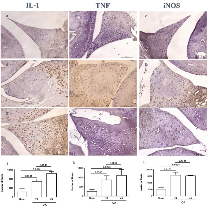
Fig 4. Representative illustration of the immunoexpression of IL-1β, TNF, and iNOS in mouse menisci subjected to postsurgical experimental OA or a sham procedure.
While mostly cells closer to the “red zone” of the meniscus stain positive in sham samples (a,b,c) there is diffuse immunoexpression in samples from OA groups sacrificed 21 (d,e,f) or 49 (g,h,i) days after surgery. Quantitation of immunoexpression from 3 independent experiments reveals significantly increased immunoexpression in OA samples, as compared to sham (j,k,l); original magnification × 400.

