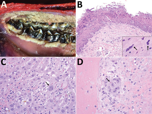
Figure 2.

Macroscopic and microscopic lesions of peste des petits ruminants virus–infected saiga, Mongolia, 2016–2017. A) Erosion and necrosis of the oral mucosa along the gingival margin of the molar teeth. B) Erosion and necrosis of the superficial oral mucosa with multifocal epithelial syncytia (inset, arrows). Original magnification ×200; inset ×1,000. C) Multifocal hepatocellular necrosis (upper and lower left, upper right) with dissolution of hepatic cords, occasional hepatocellular syncytia (black arrow), and prominent eosinophilic viral inclusion bodies, both intranuclear and chromatin (black arrow) and globular to amorphous within the cytoplasm (white arrow). Original magnification ×400. D) Bile ductule showing eosinophilic intraepithelial intracytoplasmic viral inclusion (arrow) and mild cellular degeneration with focal luminal cellular debris. Original magnification ×600.
