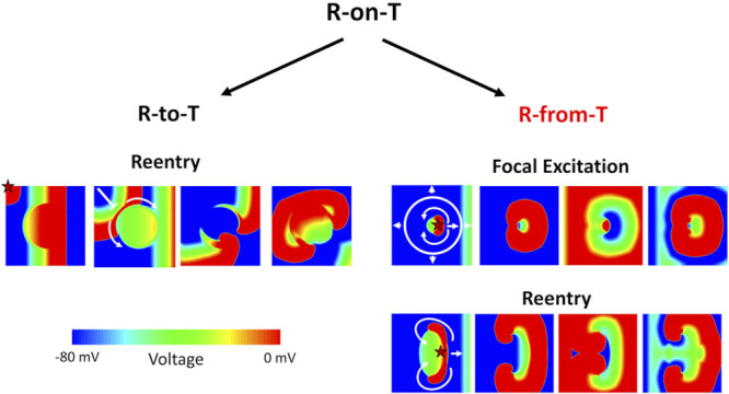
Figure 8.

Schematic diagram distinguishing the R-to-T and R-from-T mechanisms of arrhythmia initiation. Spontaneous arrhythmias that initiate with an R-on-T phenomenon on ECG can arise from 2 different underlying mechanisms, which we call R-to-T and R-from-T. A heterogenous long action potential duration (APD) region is placed in the center of the tissue. The star marks the origin of the ectopic premature ventricular complex (PVC) in each scenario. Left, R-to-T is the mechanism in which an ectopic PVC encounters a repolarizing region, undergoing unidirectional conduction block and causing figure-of-eight reentry. The R wave and T wave are 2 independent events, and the arrhythmias are only reentry. Right, R-from-T is the mechanism investigated in this study, where a PVC itself emerges from a repolarization gradient resulting in spontaneous unidirectional propagation. The R wave is caused by the T wave, and the arrhythmias can be either focal excitations (top) or reentry (bottom) depending on the properties of the heterogeneity. An animated version of this figure is shown in Movie VI in the Data Supplement.
