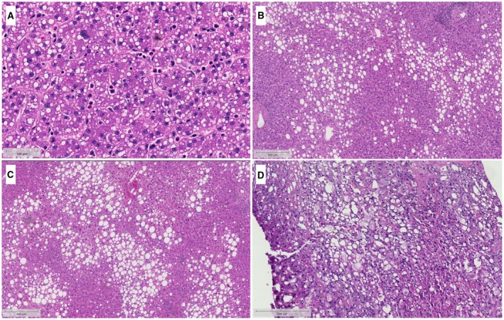
Figure 2.

Distinguishing between sd‐MaS and ld‐MaS. Diffuse sd‐MaS (A) presents as small, intracytoplasmic vacuoles that do not displace the nucleus away from its normal central location. Due to the small vacuole size, sd‐MaS can be difficult to see at lower magnification, and so this image is shown at a higher magnification than the ld‐MaS figures (B‐D). On the other hand, ld‐MaS (B‐D) presents as a large vacuole that displaces the nucleus to an eccentric location. ld‐MaS is further categorized as mild (<30%, B), moderate (30%‐60%, C), and severe (>60%, D). Courtesy Johns Hopkins Hospital Department of Pathology.
