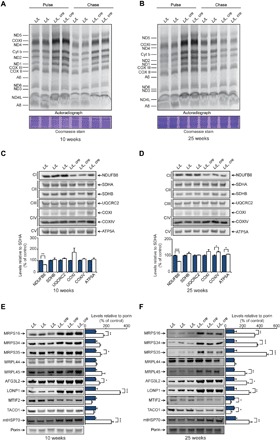
Fig. 3. Loss of MTIF3 results in uncoordinated mitochondrial protein synthesis.

(A) Levels of de novo protein synthesis were measured in heart mitochondria from control (L/L) and knockout (L/L, cre) 10-week-old mice by pulse and chase incorporation of 35S-labeled cysteine and methionine. Mitochondrial protein was separated by SDS–polyacrylamide gel electrophoresis (SDS-PAGE), stained with Coomassie, and visualized by autoradiography. Representative gels of five independent biological experiments are shown. (B) Levels of de novo protein synthesis in heart mitochondria from 25-week-old control (L/L) and knockout (L/L, cre) mice, determined as in (A). Mitochondrial proteins from isolated heart mitochondria of control (L/L) and knockout (L/L, cre) 10-week-old (C) and 25-week-old (D) mice were resolved on 4 to 20% SDS-PAGE gels and immunoblotted using antibodies to investigate the steady-state levels of OXPHOS proteins. SDHA was used as a loading control. Levels of mitoribosomal proteins, proteases, and MTIF2 proteins from isolated heart mitochondria of control (L/L) and knockout (L/L, cre) 10-week-old (E) and 25-week-old (F) mice. Porin was used as a loading control. All values are means ± SEM. *P < 0.05, **P < 0.01, ***P < 0.001, Student’s t test.
