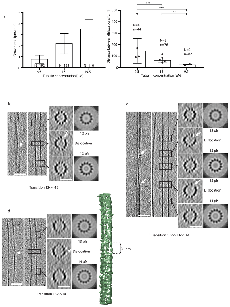
Figure 3.
Dislocation defects in the microtubule lattice detected by cryo-electron microscopy. (a) Growth rate (left) and mean distances between dislocations (right) in centrosome-nucleated microtubules as a function of the free tubulin concentration. N in the left graph denotes the number of microtubules analyzed. N and n in the right graph denote the number of cryo-electron microscopy samples analyzed and the total number of dislocations detected, respectively (see Methods and Supplementary Information). Mean±sd values for the spatial frequencies of dislocations are 0.007±0.005 μm-1, 0.016±0.005 μm-1 and 0.040±0.005 μm-1 for 6.5 μM, 13 μM and 19.5 μM free tubulin, respectively. All p-values are <0.001. (b-d) Cryo-electron tomograms of protofilament number transitions. The left panels show longitudinal slices through the microtubule in the transition region whereas the right panels show transverse sections. The left longitudinal section shows a cut through the transition region, whereas the right longitudinal section shows a cut through a central part of the microtubule. The transverse sections are averaged over the height of the black rectangles shown in the longitudinal sections (corresponding regions are indicated by black arrows) and correspond to N-fold rotational averages of the closed microtubule regions (N denotes the protofilament number). White arrows indicate the free end of a protofilament (dislocation) with the accompanying gap in the lattice at the transition. The scale bars are 50 nm for longitudinal and 25 nm for transverse sections. (b) 12⌦13 protofilament number transition. (c) 12⌦13 and 13⌦14 protofilament number transitions in close proximity in the same microtubule. (d) 13⌦14 protofilament number transition. The image to the far right shows the 3D rendering of the microtubule with the dislocation at the edge of the microtubule. A gap in the lattice with an approximate length of 31 nm is clearly visible. In (d) both longitudinal sections are identical.

