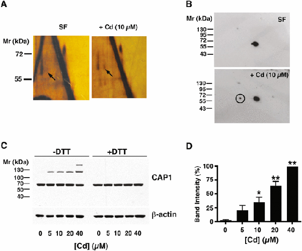
Fig. 1 – Identification of CAP1 as a disulfide-crosslinked protein in Cd-treated cells.

A) Two-dimensional electrophoresis of cell extracts of mesangial cells held in serum-free (SF) conditions for 6 h or treated with 10 μM CdCl2 under the same conditions. The gels are silver stained. Bulk proteins lie on the diagonal. Arrows indicate a spot migrating vertically off-diagonal after reduction, present in the Cd-treated cells that is absent in SF control. B) Western blot for CAP1 of gels similar to those shown in A). The major spot is on-diagonal, and the circled off-diagonal spot in the Cd-treated sample corresponds to the spot indicated by the arrow in A). The circled spot was excised and analyzed by mass spectrometry (see text). C) Western blot of CAP1 in extracts of cells treated with increasing concentrations of CdCl2, without (left side) or with (right side) dithiothreitol treatment, as indicated. A Western blot of -actin is shown as a loading control. D) The histogram to the right of the gel shows the intensity of the upper band from the -DTT cells (mean ± SD, n= 3). Values significantly different from the [Cd]=0 control calculated by ANOVA with Dunnett’s post hoc test are indicated (*, p<0.05; **, p<0.001).
