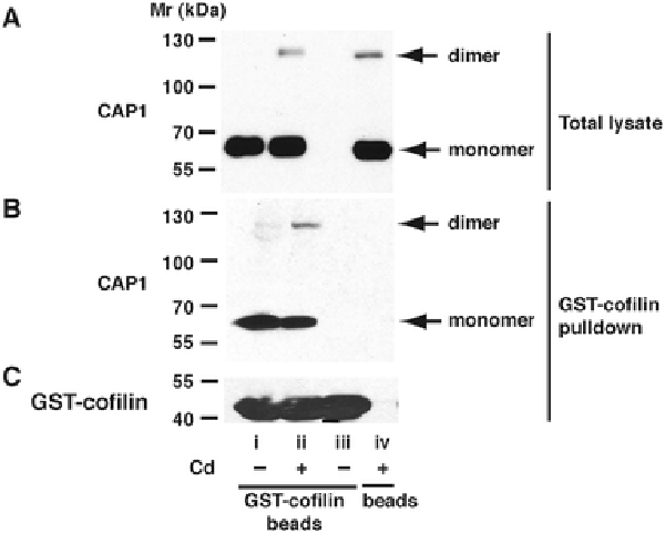
Fig. 10 – Pulldown of CAP1 by GST-cofilin from lysates of Cd2+-treated cells.

Blot A) shows the presence of CAP1 in the total lysate, with monomer and dimer bands indicated. Blot B) shows CAP1 pulled down from these lysates by GST-cofilin. Blot C) shows GST-cofilin in the same pulldowns as B). The labels under C) apply to all blots. Lane i - Lyastes from control cells, pulldown performed with GST-cofilin-conjugated beads. Lane ii - Lyastes from cells treated with 20 μM Cd2+, 6 h, pulldown performed with GST-cofilin-conjugated beads. iii) No cell lysate; GST-cofilin-conjugated beads in buffer alone. iv) Lysate of Cd2+-treated cells, pulldown procedure performed with glutathione Sepharose beads not conjugated with GST-cofilin.
