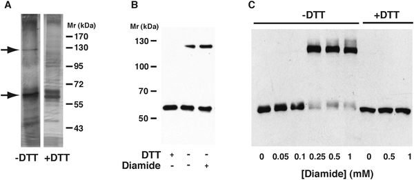
Fig. 3 – Disulfide crosslinking of recombinant CAP1.

A) Silver stained gels of recombinant CAP1 protein under non-reducing condition (-DTT) or after treatment with dithiothreitol (+DTT). Arrows indicate the positions of monomereric (lower) and putative dimeric (upper) proteins. B) Western blot with anti-CAP1 antibody of recombinant CAP1 protein, showing a higher Mr band in the absence of DTT in native protein and after treatment with diamide. Note that different Mr markers are used on this gel, and because of better linearity the Mr values reported in the text (56 kDa and 117 kDa) were calculated from this marker set. C) Western blot of CAP1 in cell extracts after treatment with increasing concentrations of diamide. The upper band (putative dimer) appears above 0.1 mM diamide, at the expense of the monomeric protein, and is sensitive to DTT (three rightmost lanes).
