
Fig. 7 – Effect of the glutathione synthesis inhibitor, BSO, on CAP1 levels.
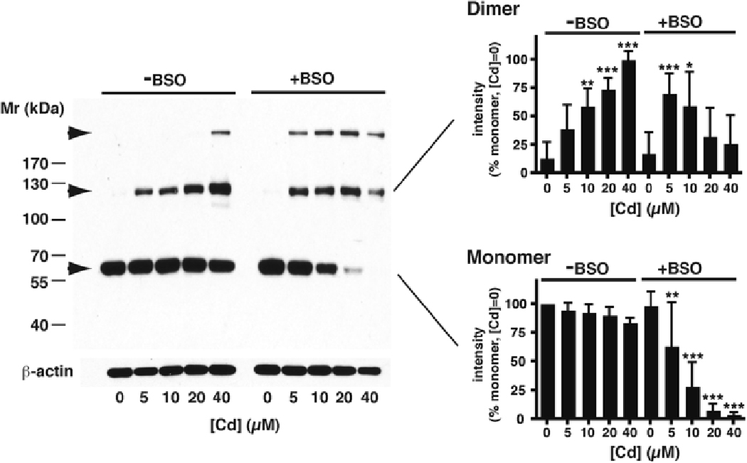
A typical Westerm blot of CAP1 is shown after treatment of cultures with increasing amounts of CdCl2 in serum-free conditions for 6 h, either without BSO (left side) or following 16 h pretreatment with BSO (right side). Arrows indicate the lower monomer band, intermediate dimer band, and a higher Mr band (upper-most arrow) discussed in the text. Blotting with anti- -actin antibody on the same gel is included as a loading control. The histograms show the mean ± SD (n=6) of intensities of the monomer and dimer bands, all normalized to the intensity of the monomer band without Cd or BSO treatment, taken as 100 %. Significant differences against the no-added Cd samples, of the same band within the same treatment group, are indicated (*, p<0.05; **, p<0.01; ***, p<0.001).