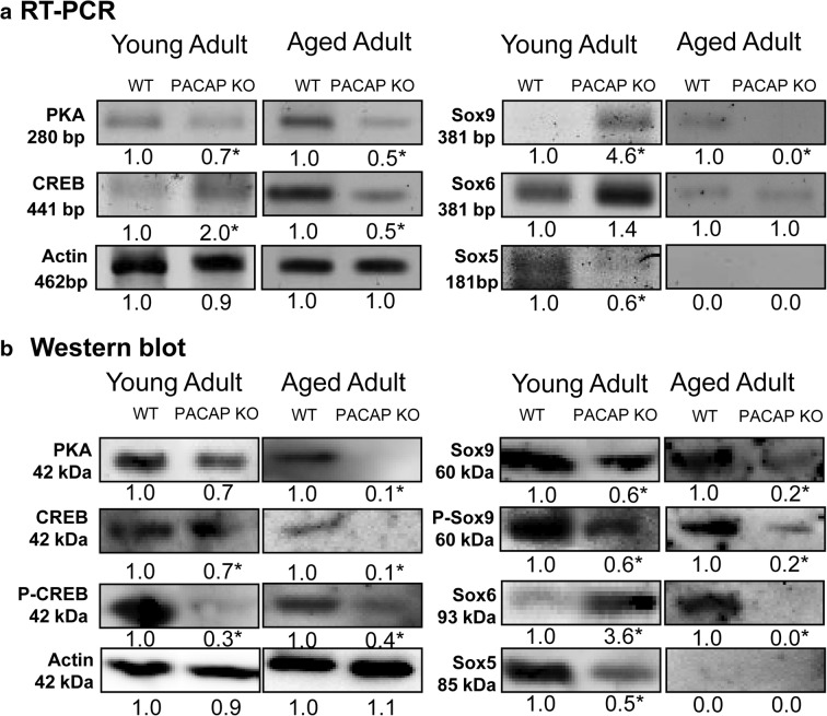
Fig. 4.
Investigation of canonical PACAP signalization in cartilage. mRNA (a) and protein (b) expression of PKA, CREB, Sox9, Sox6, and Sox5. For RT-PCR and Western blot reactions, actin was used as control. Optical signal density was measured and results were normalized to the WT controls. For a and b, numbers below signals represent integrated signal densities determined by ImageJ software. Asterisks indicate significant (*p < 0.05) alteration of expression compared to the respective control. Representative data of 5 independent experiments

