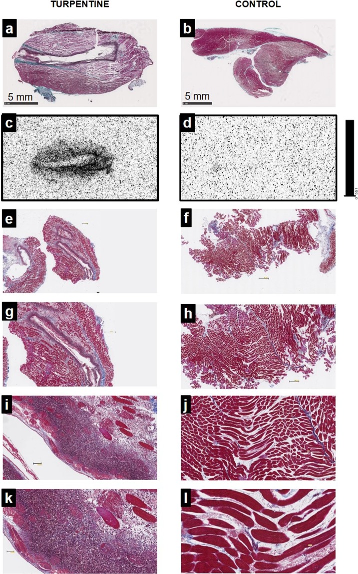
Figure 5.
Masson’s trichrome staining (a,b,e–l) and autoradiography (c,d) of gastrocnemius muscles 2 days after intramuscular injection of saline (control, left hind limb) or turpentine oil (right hind limb). Animals were sacrificed 1 hour after intravenous 99mTc-HYNIC-D-P8RI administration. Masson’s trichrome staining in (a) and (b) corresponds to the same slices of autoradiography in (c) and (d). Turpentine injected muscle (a) displays evidence of inflammation with edema and major leukocyte infiltration (e,g,i,k: x1.25, x2.5, x10, x20 respectively), co-localized with a high uptake of 99mTc-HYNIC-D-P8RI. The representative micrographs (b) corresponds to a control muscle matching with a low uptake of 99mTc-HYNIC-D-P8RI (d). Higher magnifications of the control show no evidence of inflammation (f,h,j,l).

