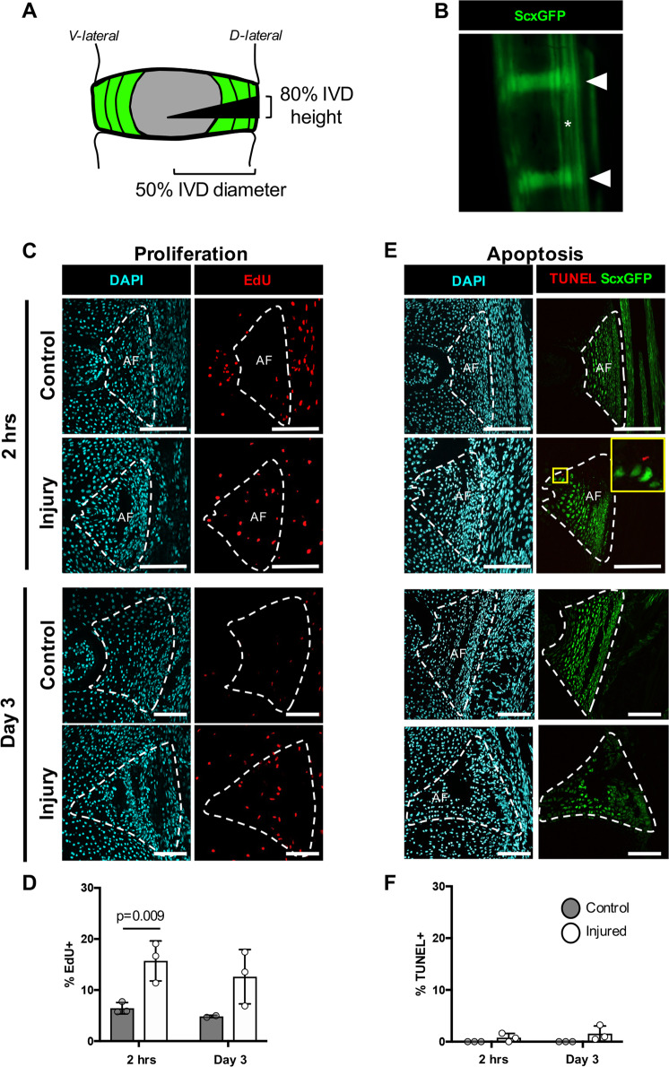
Fig. 1. Early proliferation and minimal apoptosis of injury site cells occurs immediately following neonatal herniation.
Neonatal injuries were performed using a 31G beveled syringe needle tip to 50% of the IVD diameter a in the ScxGFP reporter mouse that labels annulocytes (triangles) and tenocytes (asterisks) b. ScxGFP expression is decreased in the neonatal IVD 2 hrs post-injury (yellow triangle) b. Proliferating cells were detected using EdU. Representative images of the posterior control AF of an uninjured IVD and the posterior AF of an injured IVD show an increase in proliferation at 2 hrs and d3 c. Quantification using cell counting determined that there was a significant increase in the percentage of proliferating cells at d0 (2 hrs post-injury) compared to controls d. Cells undergoing apoptosis were detected using TUNEL staining. Minimal apoptosis was observed in uninjured controls at 2 hrs, where the few cells stained positive for TUNEL were located at the border of ScxGFP+ cells and ScxGFP- cells in the injured AF and at d3 e. No differences between the percentage of TUNEL-positive cells were observed between control and injured AFs f. Error bars = SD. Scale = 100 μm.

