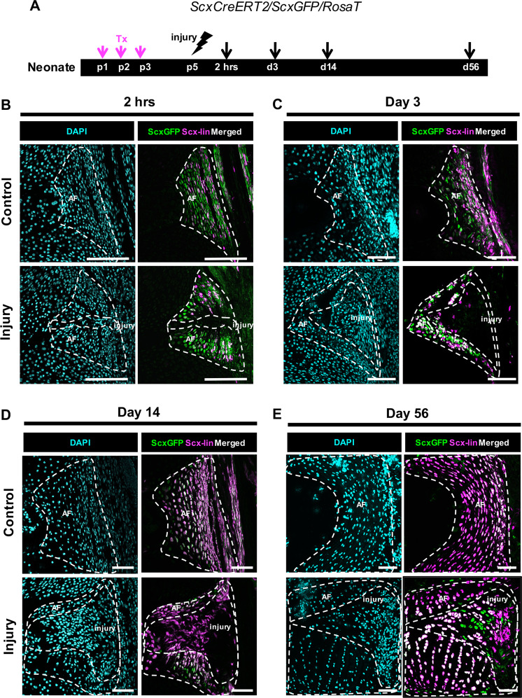
Fig. 4. Neonatal AF healing occurs via a transient loss of ScxGFP in Scx-lin annulocytes followed by restoration of ScxGFP.
ScxCreERT2/RosaT/ScxGFP mice were used to trace the fate of annulocytes that were labeled with tamoxifen at p1-p3, and traced after injury at 2 hrs, d3, d14, and d56 a. Scx-lin cells in the uninjured control AF were more numerous over time, with a smaller portion of annulocytes labeled at 2 hrs b compared to d56 where most annulocytes were Scx-lin e. At 2 hrs, the injury site was cellular but cells immediately adjacent to the puncture tract lost ScxGFP expression and were not Scx-lin b. At d3, the injury site expanded and contained cells that were neither ScxGFP+ nor Scx-lin c. At d14, the injury site remained ScxGFP-, and was occupied by Scx-lin cells recruited to the injury site d. By d56, differentiation of Scx-lin cells was observed by co-localization with ScxGFP e. Images were acquired at 20X and digitally magnified to show the entire posterior AF at each timepoint. Since IVDs are growing at these early stages, the length of the scale bar is decreasing with increasing timepoint. Scale = 100 μm.

