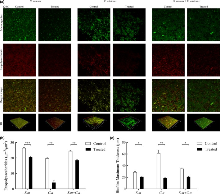
Figure 3.

Effect of CUR on the EPS of mono‐ and dual‐species biofilms by CLSM. After incubation and staining, images of the biofilms were collected. The green channel was used for microorganism (a). The red channel was used for EPS (a). The EPS and thickness of the biofilms were quantified and compared (b, c). The asterisks (*) indicate significant differences (*p < .05; **p < .01; ***p < .001)
