Figure 1.
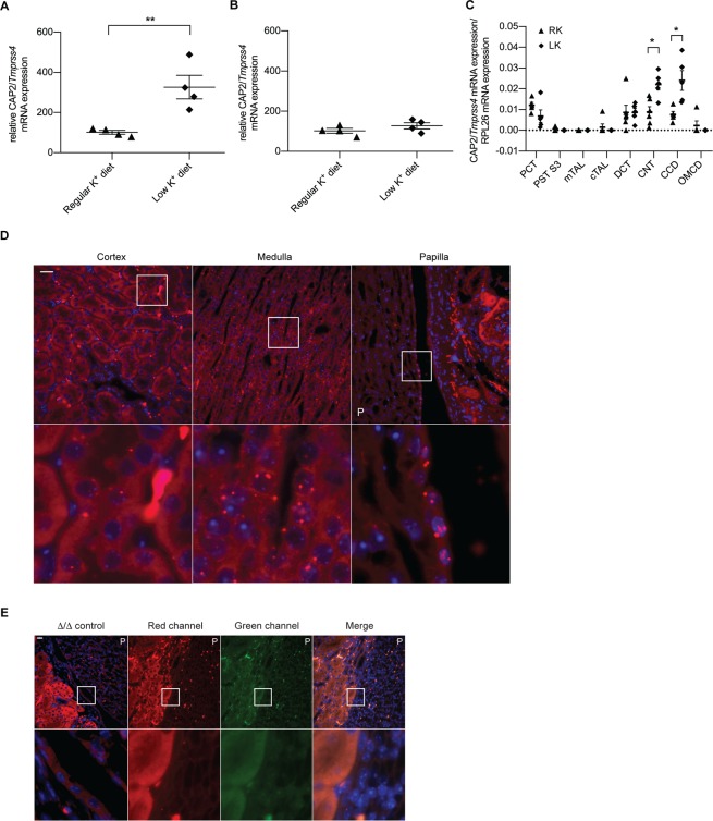
CAP2/Tmprss4 mRNA expression is upregulated by low dietary K+ in distal tubules, and also localizes to the papillary transitional epithelium. Relative mRNA transcript expression levels of CAP2/Tmprss4 in (A) kidney, and (B) colon from wildtype mice under regular K+ diet (n = 4, triangles) and low K+ diet (n = 4, diamonds). (C) Detection of wildtype CAP2/Tmprss4 mRNA transcript expression in microdissected nephron segments (n = 4–6/segment) on regular (RK) and low (LK) potassium diet. PCT: proximal convoluted tubule, PST S3: proximal straight tubule segment 3, mTAL: medullary thick ascending limb of Henle’s loop, cTAL: cortical thick ascending limb of Henle’s loop, DCT: distal convoluted tubule, CNT: connecting tubule, CCD: cortical collecting duct, OMCD: outer medullary collecting duct. (D) RNAscope detection of CAP2/Tmprss4 in renal cortex, medulla and papilla of wildtype mice following LK diet. (E) Negative control for CAP2/Tmprss4 RNAscope detection in knockout (∆/∆) kidney under low K+ diet, and negative control for RNAscope fluorescent channels (including channels shown in Fig. 3E,F) in kidney sections from wildtype mice under low K+ diet. Magnification 40×, the white boxes indicate zones of higher (63×) magnification, “P” indicates the location of the renal pyramid, scale bar represents 25 µm. * p < 0.05, **p < 0.01.

