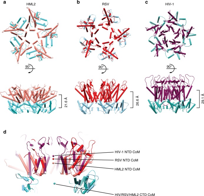
Fig. 3. Comparison of HIV-1, RSV and HML2 CA pentamers.
a–c Top and side view cartoon representations of CA pentamers with α-helices shown as cylinders, from a HML2 CArec, b RSV (built from PDB: 1EM9 and 3G1I docked into EMD 5772) and c HIV-1 (PDB: 5MCY). NTDs are coloured pink, red and purple and CTDs are coloured light cyan, light blue and cyan for HML2, RSV and HIV-1, respectively. Pentamers are aligned with respect to their CTDs. In the lower panels, the distance between the centre of mass (CoM) for each NTD and CTD ring is shown on the right. d Central section through CA pentamers aligned with respect to CTDs. The CoM for NTD and CTD rings is represented by the spheres, colour coded as in a–c and shows the coincident CoM for CTDs and the difference in CTD–NTD vertical displacement for each pentamer.

