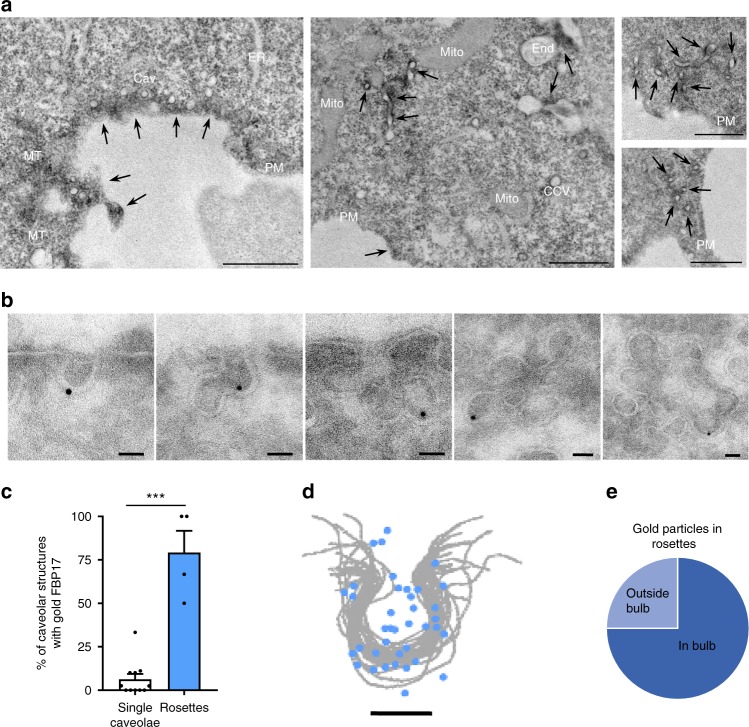
Fig. 2. FBP17 is localized in caveolae and caveolar rosettes.
a EM of cells expressing GFP-FBP17. FBP17 signal (marked with arrows) is mostly located around caveolar clusters. Arrows indicate the presence of GFP-FBP17. Mito mitochondria, PM plasma membrane, Cav caveolae, End endosome, ER endoplasmic reticulum, MT microtubules, CCV clathrin-coated vesicle. Scale 1 μm. b Localization of endogenous FBP17 by I-EM. FBP17 (gold signal) in caveolae and in rosettes with different caveolar density are shown. Scale bar 50 nm. c Quantification of the fraction of caveolar structures with gold signal associated with FBP17 antibody. N = 11 (left) and 4 (right) biologically independent cells from 1 experiment. Data represent mean ± S.E.M. Statistical analysis with a two-tailed unpaired t test. ***P < 0.005. d Localization of gold particles associated with endogenous FBP17 in caveolae. Scale bar 50 nm. e Fraction of gold particles associated with endogenous FBP17 in the indicated regions of rosettes.

