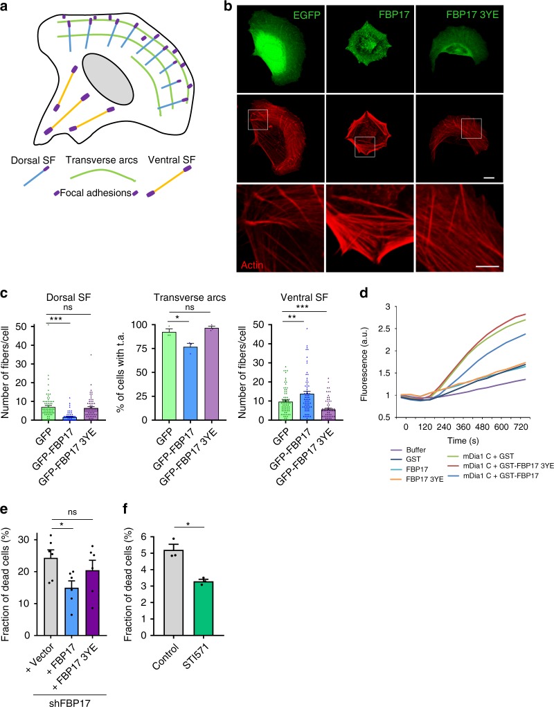
Fig. 8. PM integrity and stress fiber inhibition are regulated by tension-regulated FBP17 phosphorylation.
a Cartoon depicting the different pools of stress fibers (SF). b Cells were transfected with plasmids expressing the indicated proteins and actin was stained. Scale bar 10 μm, boxes 5 μm. c Quantification of the different stress fiber pools in each condition as in b. N = 74 (GFP), n = 68 (GFP-FBP17), and n = 68 (GFP-FBP17 3YE) biologically independent cells from 3 independent experiments, for the quantification of dorsal and ventral SF; and n = 3 biologically independent samples from 3 independent experiments for the quantification of transverse arcs (t.a.). Statistical analysis with a two-tailed unpaired t test. *P < 0.05; **P < 0.01; ***P < 0.005. d In vitro actin-pyrene polymerization regulated by mDia1C. Different proteins were incubated with mDia1C at 75 nM. Representative of three independent experiments. e FBP17 expression was silenced in human fibroblast and cells were treated with hypo-osmotic medium. Trypan blue-labeled cells were scored and the fraction of dead cells positive for trypan blue were calculated for each condition. N = 6 biologically independent samples from 6 independent experiments. Statistical analysis with a two-tailed unpaired t test. *P < 0.05; ns non-significant. f Effect of inhibiting Abl kinases in cell sensitivity to osmotic shock. Cells were pretreated with vehicle or Abl inhibitor (STI571) for 30 min and then treated with osmotic shock (30 mOsm) for 10 min and dead cells were counted in each condition. N = 3 biologically independent samples from 3 independent experiments. Statistical analysis with a two-tailed unpaired t test. *P < 0.05. Data represent mean ± S.E.M.

