Figure 3.

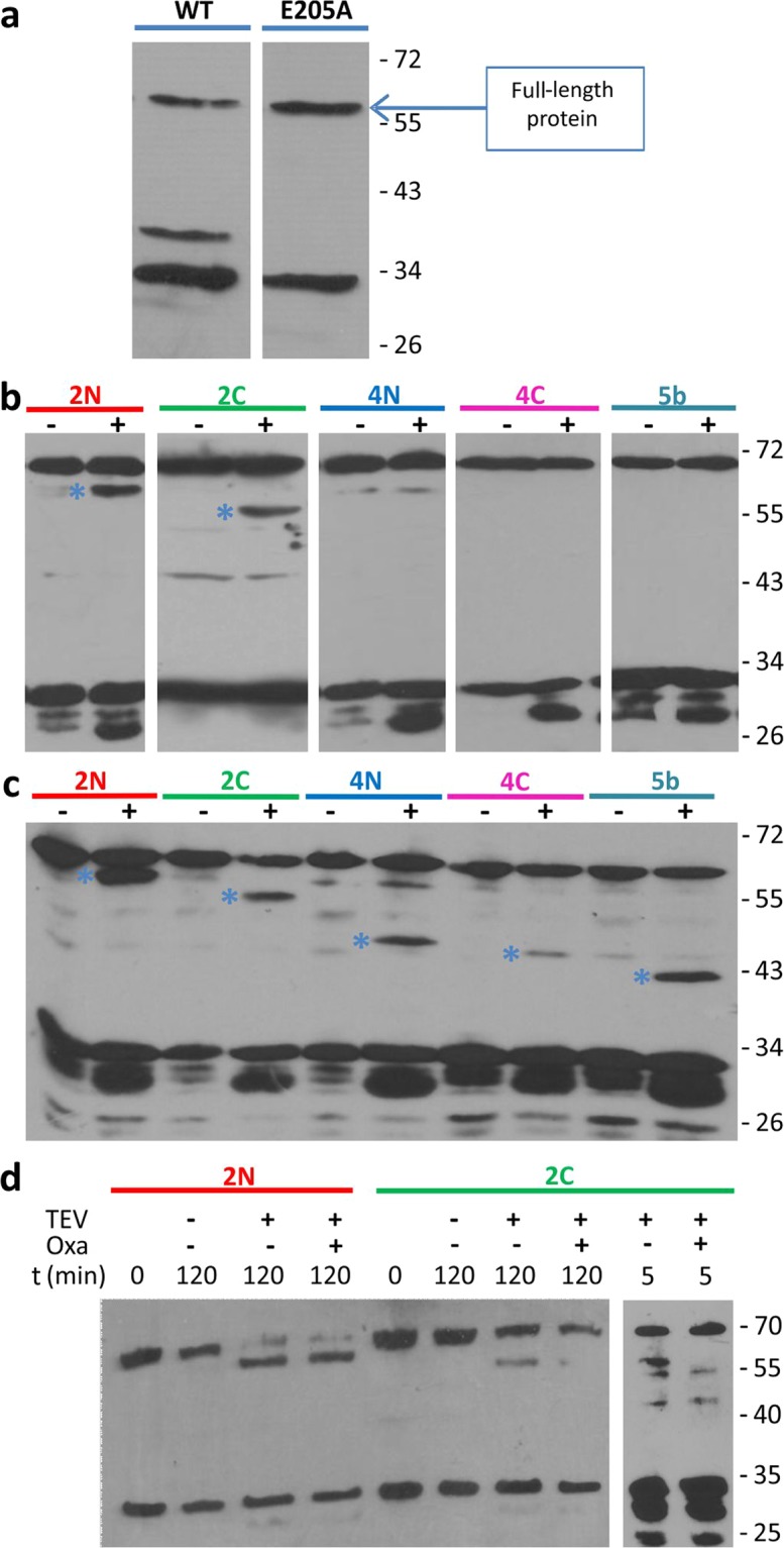
TEV Protease susceptibility assays of the MecR1.E205A.TEV insertional mutant proteins. (a) Expression of full-length MecR1 wild type and the E205A mutant (with a C-terminal His-tag) in E. coli BL21 Star (DE3), evaluated by Western blot using a His-tag specific HRP-conjugated antibody. Full, uncut gel images are provided in Fig. S13. (b) Spheroplasts expressing each MecR1.E205A.TEV insertional mutant protein were incubated in the absence (−) or presence (+) of TEV Protease, in order to determine the intra- or extracellular location of the TEV recognition peptide. (c) Membrane-protein extracts of E. coli expressing each MecR1.E205A.TEV insertional mutant protein were prepared and incubated in the absence (−) or presence (+) of TEV protease. In (b,c), the blue asterisk indicates the C-terminal fragment resulting from TEV proteolysis. The expected molecular weight of the full-length MecR1.E205A.TEV versions is 70.5 kDa. The expected molecular weight of the C-terminal His-tagged fragment after TEV treatment for each variant is: 2N, 61.3 kDa; 2C, 58.2 kDa; 4N, 47.3 kDa; 4C, 45.3 kDa and 5b, 40.1 kDa. (d) Spheroplasts expressing MecR1-TEV-2N and 2C were incubated in the presence (+) or absence (−) of oxacillin, prior to or simultaneously with TEV protease treatment. Lanes 2-4 and 6-8: incubation with oxacillin followed by incubation with 750 μg/ml TEV protease, 500 rpm. Lanes 9 and 10: simultaneous incubation with 100 μg/ml TEV protease, without agitation, as in B. Western blots were carried out using a His-tag specific HRP-conjugated antibody. Full, uncut gel images for B and D are provided in Figs. S14 and S15, respectively. Numbers on the right indicate the positions of migration of the molecular weight markers (kDa).
