
Figure 1. The intestinal microbiota play opposing roles in regulating proximal gut versus distal gut MNV infection.
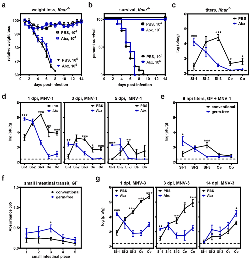
a-b) Groups of Ifnar−/− mice were treated with PBS or Abx and then infected with 105 pfu MNV-1 (n = 19 for PBS, n= 17 for Abx) or 104 TCID50 units MNV-1 (n = 4 for both groups) and followed for weight loss (a) and survival (b). c) Groups of Ifnar−/− mice (n = 7) were treated with PBS or Abx and then infected with 105TCID50 units MNV-1. At 1 day post-infection (dpi), virus titers were determined in the indicated segments of the intestinal tract. d) Groups of PBS- and Abx-treated wild-type B6 mice were inoculated with 107TCID50 units MNV-1 and virus titers determined at 1 (n = 10 for PBS, n = 8 for Abx), 3 (n = 8 for PBS, n = 6 for Abx), and 5 dpi (n = 3 per group). Groups were also tested at 14 dpi but no virus was detected. e) Conventional (n = 13) or germ-free mice (n = 15) were inoculated with 106 pfu neutral red-labeled MNV-1. At 9 hours post-infection (hpi), tissues were collected and plaque assays were used to measure newly synthesized virus. f) Conventional (n = 5) or germ-free (n = 4) mice were administered Evan’s blue dye and intestinal transit times were determined 15 min later. g) Groups of PBS- and Abx-treated wild-type B6 mice were inoculated with 107TCID50 units MNV-3 and virus titers determined at 1 (n = 7 for PBS, n = 8 for Abx), 3 (n = 6 per group), and 14 (n = 6 per group) dpi. Error bars indicate the mean of all data points. Unpaired two-tailed Student’s t-tests were used for statistical purposes. *P < 0.05, **P < 0.01, ***P < 0.001. See Supplementary Table 1 for detailed statistical information.